
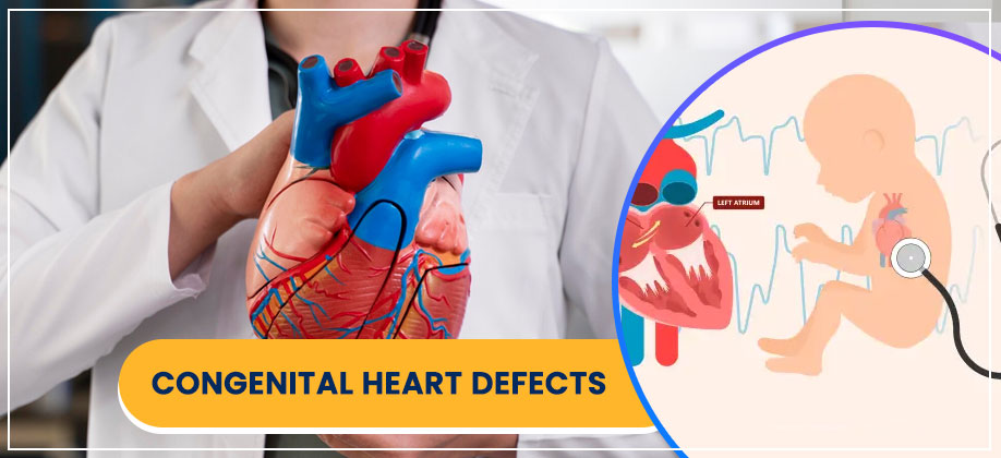
Congenital Heart Defects
Congenital Heart Defects Treatments:
Looking for the best cardiologist in Howrah for ASD closure in adults? Our team of the best child heart doctors in Howrah offers specialized care for all congenital heart defects.
Congenital Heart Defects (CHDs) are structural problems with the heart that are present at birth.They can range from mild to severe and affect how blood flows through the heart and to the rest of the body. Here’s detailed information on the common CHDs you listed:
1. Atrial Septal Defect (ASD)
- What it is: An ASD is a “hole in the heart” in the wall (septum) that separates the two upper chambers of the heart (atria). During fetal development, there are openings in this wall that usually close shortly after birth. If one remains open, it’s an ASD.
- How it affects blood flow: Oxygen-rich blood from the left atrium can flow through the hole into the right atrium, mixing with oxygen-poor blood. This extra blood then gets pumped to the lungs, making the right side of the heart and the lungs work harder.
- Symptoms: Many small ASDs are asymptomatic and may close on their own. Larger ASDs can cause:
- Heart murmur (a whooshing sound heard with a stethoscope)
- Shortness of breath, especially with activity
- Tiring easily, especially during feeding in infants
- Frequent respiratory or lung infections
- Skipped heartbeats or palpitations
- Swelling of legs, feet, or abdomen (in adulthood, if untreated)
- Diagnosis: Often detected by a heart murmur during a physical exam. Confirmed with an echocardiogram (ultrasound of the heart).
- Treatment: Small ASDs may be monitored as they can close spontaneously.Larger defects that cause symptoms or risk complications (like pulmonary hypertension, heart failure, or stroke) may require closure, either via a catheter-based procedure (device closure) or open-heart surgery.Child heart doctors have special expertise in doing complex and borderline ASD device closures.
2. Ventricular Septal Defect (VSD)
- What it is: A VSD is a hole in the wall (septum) that separates the two lower chambers of the heart (ventricles). It’s one of the most common CHDs.
- How it affects blood flow: Oxygen-rich blood from the left ventricle can flow through the hole into the right ventricle, mixing with oxygen-poor blood and being pumped back to the lungs. This increases blood flow to the lungs and makes the heart work harder.
- Types: VSDs can occur in different parts of the ventricular septum, including perimembranous (most common), muscular, inlet, and conoventricular.
- Symptoms: Symptoms depend on the size of the hole.Small VSDs may be asymptomatic and close on their own. Larger VSDs can cause:
- Heart murmur (often loud)
- Shortness of breath or rapid breathing
- Difficulty feeding and poor weight gain (failure to thrive in infants)
- Sweating during feeding
- Frequent respiratory infections
- Fast heart rate
- Diagnosis: Often detected by a heart murmur. Confirmed by echocardiogram.
- Treatment: Small VSDs often close spontaneously. Larger VSDs causing symptoms or complications (like heart failure, pulmonary hypertension) may require medications to manage symptoms and/or surgical closure (patching the hole) or catheter-based closure. child heart doctors team are the only team to do maximum borderline VSD device closure(VSDs at difficult location like under the aortic or pulmonary valve)
3. Patent Ductus Arteriosus (PDA)
- What it is: The ductusarteriosus is a blood vessel that connects the aorta (the main artery carrying oxygen-rich blood to the body) to the pulmonary artery (which carries oxygen-poor blood to the lungs) in a fetus. This connection allows blood to bypass the developing lungs. Normally, it closes within a few days after birth. If it remains open, it’s called a patent (open) ductusarteriosus.
- How it affects blood flow: After birth, if the PDA remains open, oxygen-rich blood from the aorta flows back into the pulmonary artery, sending extra blood to the lungs. This can overwork the lungs and the left side of the heart.
- Symptoms: Small PDAs may be asymptomatic. Larger PDAs, especially in premature babies, can cause:
- Heart murmur (a continuous “machinery-like” murmur)
- Rapid breathing or shortness of breath
- Poor feeding and poor weight gain
- Sweating with crying or eating
- Easy tiring
- Frequent lung infections
- Diagnosis: Detected by a characteristic heart murmur. Confirmed with an echocardiogram.
- Treatment: In premature infants, medications (like NSAIDs, e.g., indomethacin) can sometimes help close the PDA. If not, or in full-term infants and children, it may be closed via a catheter-based procedure (coil or device embolization) or surgical ligation (tying off the vessel).Child Heart Doctors are the first team to do a preterm PDA device closure in a 700 gm baby who was successfully discharged.
4. Aortopulmonary Window (AP Window)
- What it is: A rare congenital heart defect characterized by a direct abnormal connection (hole) between the aorta and the main pulmonary artery, just above the heart. It results from incomplete separation of these two major vessels during fetal development.
- How it affects blood flow: Similar to a large PDA or VSD, an AP window allows oxygen-rich blood from the high-pressure aorta to shunt into the lower-pressure pulmonary artery, leading to excessive blood flow to the lungs and increasing pressure in the pulmonary arteries.
- Symptoms: Symptoms are usually present early in infancy and are due to heart failure:
- Rapid breathing (tachypnea)
- Poor feeding and delayed growth (failure to thrive)
- Sweating (diaphoresis)
- Irritability
- Rapid heartbeat
- Recurrent respiratory infections
- Diagnosis: Often suspected based on physical exam findings (heart murmur, signs of heart failure). Confirmed with echocardiogram, and sometimes cardiac MRI or CT scan for more detailed anatomy.
- Treatment: Surgical repair is necessary in few cases and should be performed as soon as possible after diagnosis to prevent irreversible damage to the lung blood vessels (pulmonary hypertension). The hole is typically patched. In present era AP Window is device closed in maximum cases which avoids surgery.
5. Tetralogy of Fallot (TOF)
- What it is: A complex congenital heart defect involving four specific abnormalities that occur together:
- Ventricular Septal Defect (VSD): A hole between the lower heart chambers.
- Pulmonary Stenosis: Narrowing of the pulmonary valve and/or artery, restricting blood flow to the lungs.
- Overriding Aorta: The aorta is positioned directly over the VSD, receiving blood from both ventricles instead of just the left.
- Right Ventricular Hypertrophy: The muscle wall of the right ventricle thickens due to the increased workload of pumping against the narrowed pulmonary artery.
- How it affects blood flow: Due to pulmonary stenosis, blood flow to the lungs is reduced.The VSD and overriding aorta allow oxygen-poor blood from the right ventricle to mix with oxygen-rich blood in the left ventricle and be pumped to the body.This leads to reduced oxygen levels in the systemic circulation.
- Symptoms: The primary symptom is cyanosis (bluish discoloration of the skin, lips, and nail beds) due to low oxygen levels in the blood. Other symptoms include:
- “Tet spells”: Sudden, severe episodes of increased cyanosis, hyperpnea (rapid, deep breathing), and irritability, often triggered by crying or feeding. Infants may instinctually squat to relieve these spells.
- Heart murmur
- Tiring easily, especially during feeding
- Clubbing of fingers and toes (in older children)
- Fainting or seizures (during severe tet spells)
- Diagnosis: Often suspected prenatally or at birth due to cyanosis or a heart murmur. Confirmed with echocardiogram. Chest X-ray and ECG may also be used.
- Treatment: Surgical repair is typically required, usually in infancy.
- Complete repair: Involves closing the VSD with a patch and widening the pulmonary valve and/or artery to improve blood flow to the lungs.
- Palliative surgery (shunt): In some cases, a temporary shunt may be placed (e.g., Blalock-Taussig shunt) to increase blood flow to the lungs if the baby is too small or sick for complete repair. Complete repair is then performed later.
- In present era palliative RVOT stent is done or PDA stenting done to increase blood flow to lungs.Childheart doctors team is known to do maximum no of RVOT stents in eastern zone in both preoperative and postoperative cases.
6. Transposition of the Great Arteries (TGA)
- What it is: A serious and critical CHD where the two main arteries leaving the heart are switched (transposed):
- The aorta arises from the right ventricle (instead of the left).
- The pulmonary artery arises from the left ventricle (instead of the right).
- Types:
- D-transposition of the great arteries (d-TGA): The most common type, where the ventricles are in their normal positions, but the arteries are switched. This creates two separate, parallel circulatory systems, leading to severe cyanosis immediately after birth as oxygen-poor blood is recirculated to the body and oxygen-rich blood to the lungs. Survival depends on mixing between the two circulations (e.g., via an ASD or PDA).
- L-transposition of the great arteries (l-TGA or congenitally corrected TGA): A less common type where the ventricles are also switched in addition to the arteries (the right ventricle is on the left, and the left ventricle is on the right). In this case, blood flow can sometimes be “corrected” functionally, but the right ventricle is performing the systemic circulation, which can lead to long-term issues like heart failure.
- Symptoms (d-TGA): Severe cyanosis (blue or gray skin, lips) immediately after birth, poor feeding, rapid breathing, and weak pulse. Without immediate intervention, it is life-threatening.
- Symptoms (l-TGA): May not be apparent until later in childhood or adulthood, or can present with heart failure or heart block.
- Diagnosis: Often diagnosed prenatally via fetal echocardiogram or soon after birth due to severe cyanosis. Confirmed by echocardiogram.
- Treatment (d-TGA):
- Prostaglandin E1 (PGE1) infusion: Given immediately after birth to keep the PDA open, allowing some mixing of blood.
- Balloon atrial septostomy (Rashkind procedure): A catheter-based procedure performed to enlarge an existing ASD or create a new one, improving blood mixing.
- Arterial switch operation (Jatene procedure): The definitive surgical repair, typically performed within the first few weeks of life, where the great arteries are switched back to their correct ventricles.
- Treatment (l-TGA): Management depends on associated defects and symptoms, often involving close monitoring and sometimes surgical repair of associated lesions.
7. Total Anomalous Pulmonary Venous Connection (TAPVC)
- What it is: A rare and critical CHD where the four pulmonary veins (which normally bring oxygen-rich blood from the lungs to the left atrium) connect abnormally to the right side of the heart instead.
- How it affects blood flow: All oxygen-rich blood from the lungs returns to the right atrium, where it mixes with oxygen-poor blood. This mixed blood then flows to the left side of the heart (usually through an ASD, which is essential for survival in this condition) and is pumped to the body. However, the body receives less oxygen than it needs, and there’s excessive blood flow to the right side of the heart and lungs.
- Types: TAPVC can be classified based on where the pulmonary veins connect to the right side of the heart (e.g., supracardiac, cardiac, infracardiac, or mixed). Some types can also involve obstruction to the venous return, making it more critical.
- Symptoms: Symptoms are usually present from birth or shortly after, and include:
- Cyanosis (bluish skin color)
- Rapid and difficult breathing
- Poor feeding
- Lethargy/extreme sleepiness
- Weak pulse
- Diagnosis: Often suspected based on clinical signs and chest X-ray. Confirmed with echocardiogram, which visualizes the abnormal venous connections.
- Treatment: Surgical repair is emergent, especially if there is obstruction to pulmonary venous return. The goal is to reconnect the pulmonary veins to the left atrium and close any abnormal connections or shunts (like the ASD).
- Palliative verical vein balloon dilatation or stenting is done in critically ill neonates with severe obstruction.
8. Coarctation of the Aorta (COARCTATION)
- What it is: A narrowing of the aorta, the main artery that carries oxygen-rich blood from the heart to the body. It most commonly occurs in the segment of the aorta just beyond where the arteries to the head and arms branch off (the aortic arch).
- How it affects blood flow: The narrowing restricts blood flow to the lower part of the body. This forces the left ventricle to pump harder to push blood through the narrowed aorta, leading to high blood pressure in the upper body and head, and lower blood pressure in the legs and abdomen.
- Symptoms: Symptoms depend on the severity of the narrowing:
- Severe coarctation in newborns: Can lead to signs of heart failure or shock shortly after birth, including difficulty breathing, poor feeding, irritability, and pale skin.
- Milder coarctation (later in childhood or adulthood): May be asymptomatic or cause:
- High blood pressure in the arms (often a difference in blood pressure between arms and legs)
- Weak or absent pulses in the legs
- Leg cramps with exercise
- Headaches
- Nosebleeds
- Chest pain
- Dizziness or fainting
- Diagnosis: Often detected during a physical exam by a difference in pulses or blood pressure between the upper and lower extremities. A heart murmur may also be present. Confirmed with echocardiogram, and sometimes cardiac MRI or CT scan for precise anatomical details.
- Treatment:
- Surgery: The narrowed segment of the aorta is removed or widened.This can involve end-to-end anastomosis (reconnecting the two healthy ends) or using a patch.
- Balloon angioplasty with or without stent placement: A catheter-based procedure where a balloon is inflated to open the narrowed area, and sometimes a stent is placed to keep it open.
- Prognosis: Surgical repair generally has excellent outcomes, but lifelong follow-up is necessary due to the risk of re-coarctation, hypertension, or aneurysm formation.
These congenital heart defects represent a significant health challenge, and early diagnosis and appropriate management are crucial for improving outcomes and quality of life for affected children.
FAQs
- Q1: Can CHDs be detected before birth?
Yes. Many CHDs are detected during routine prenatal scans. Fetal echocardiograms provide a detailed look at the baby’s heart. - Q2: Will my child need surgery for every CHD?
Not always. Some defects are mild and close on their own. Others may require catheter procedures or surgery. - Q3: How safe is heart surgery for infants?
Pediatric heart surgery is highly advanced and safe when performed by experienced teams. - Q4: Will my child need lifelong follow-up?
Yes, regular heart check-ups are essential, even after successful treatment, to ensure lifelong health. - Q5: Can my child live a normal life after treatment?
Most children with treated CHDs grow up to lead healthy, active lives with little or no restrictions.
All Services:
- Fetal Cardiology
- Pediatric Arrhythmia Management
- Interventional Cardiology
- Cardiac Imaging Service
- Congenital Heart Defects
- Acquired Heart Conditions
- Symptoms & Diagnosis of Heart Conditions in Children
- Congenital Muscular Dystrophy (CMD)
- Heart Failure in Children
- Pulmonary Arterial Hypertension (PAH)
- Congenital Heart Disease
- Special Heart Tests for Children
- Special Heart Interventions for Children
For Appoinment Call
(+91) 91630 48066